The loss of motor units with age is an inevitable process: What is it and how do we measure it?1/23/2024 By Avery Hinks In my previous knowledge translation articles, I’ve given extensive attention to the loss of muscle mass and strength with age. I’ve talked less, however, about how that loss comes about. When discussing those losses in muscle mass and function, one can arguably point to one aspect of the neuromuscular system: the motor unit. What is a motor unit? Any conscious muscle action—whether we’re lifting a cup of coffee, kicking a soccer ball, or doing a backflip—begins with an electrical signal from the brain. That signal will travel from the brain down the spinal cord to something called an alpha motor neuron. Each alpha motor neuron connects through a nerve to a particular set of muscle fibres within a muscle. The signal will pass through the alpha motor neuron, down the nerve, then cause the muscle fibres associated with that motor neuron to contract. An alpha motor neuron together with the muscle fibres it controls is called a motor unit, and this is the smallest anatomical unit under voluntary control. There are hundreds of motor units in many muscles of the human body! 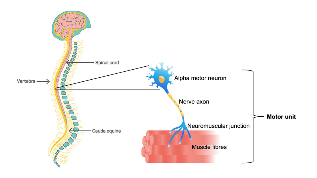 The number and type of motor units recruited depends on the action being performed. Lighter, more finely controlled tasks like lifting a coffee cup require motor units that activate “slow twitch” muscle fibres, which produce a small amount of force and contract slowly. These motor units require a lower electrical signal from the brain to activate, labeled as “low threshold.” Lifting something heavier—say you’re helping a friend move their desk—will require motor units that activate “fast twitch” muscle fibres, which produce more force and contract fast. These motor units are “high threshold,” activated when a stronger electrical signal is sent from the brain. Regardless of the task, motor units will be recruited in order of lowest threshold to highest threshold. What happens to motor units throughout the lifespan?Throughout adulthood, motor units go through cycles of denervation and reinnervation. Essentially, the connection between an alpha motor neuron and its muscle fibres is severed then rebuilt anew, again and again. When that process is healthy, it looks something like the figure below. Note that the fibres are reinnervated by the same alpha motor neuron that originally innervated them. 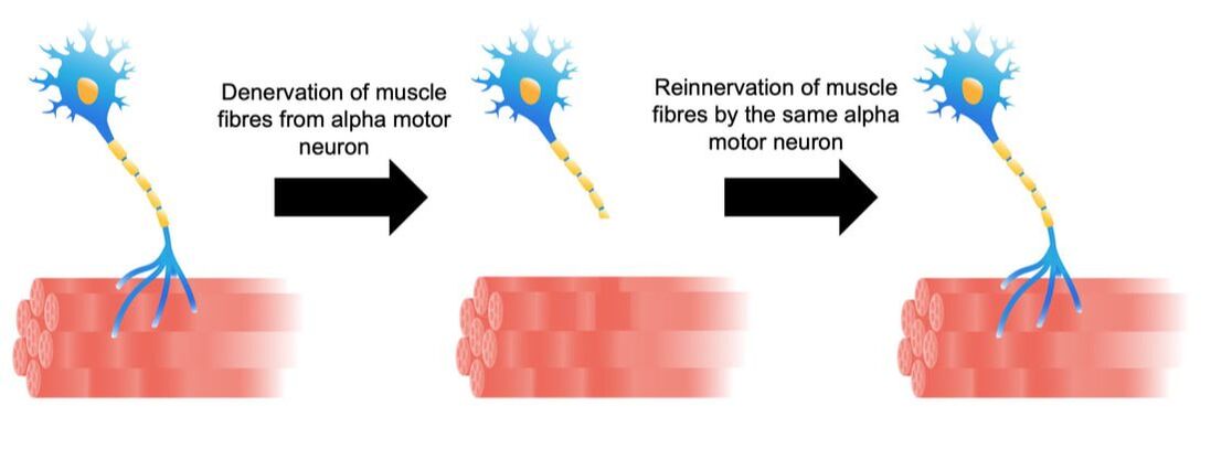 As aging progresses, this process changes form. Sometimes, the alpha motor neuron dies, and its muscle fibres are instead added to that of a neighbouring motor unit. These fibres then take on the fibre type of their new alpha motor neuron. For example, if the fibres were previously fast twitch, but became innervated by a low-threshold motor neuron, they would transition to slow twitch. Sometimes, however, that doesn’t even happen. Sometimes those leftover muscle fibres are not reinnervated by anything, and wither away. 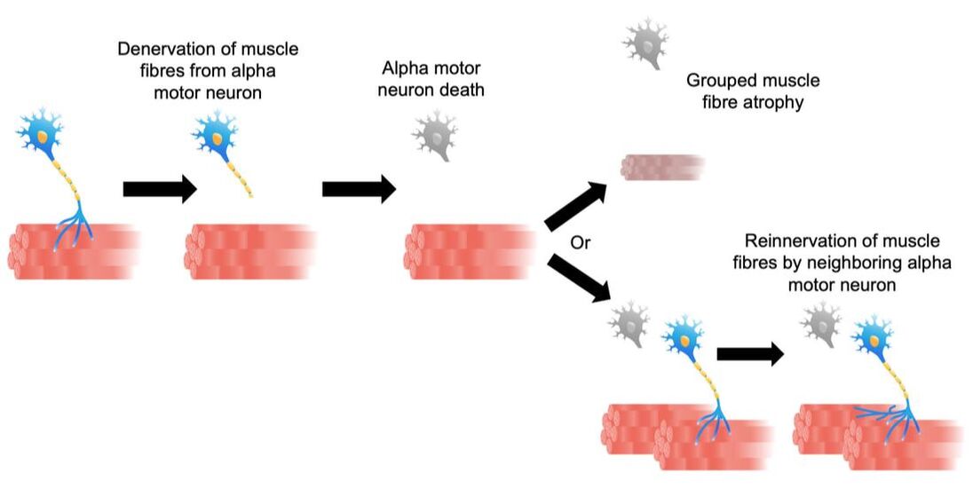 How is motor unit loss measured?A motor unit’s activity manifests as an electrical signal called an action potential. Researchers can detect action potentials using a technique called EMG, which involves placing electrodes over the muscle of interest. These can be surface electrodes, which are like stickers and pick up activity of many motor units at once, or needle electrodes, which are inserted through the skin to pick up activity of a single motor unit. To estimate total motor unit number, researchers need two values:
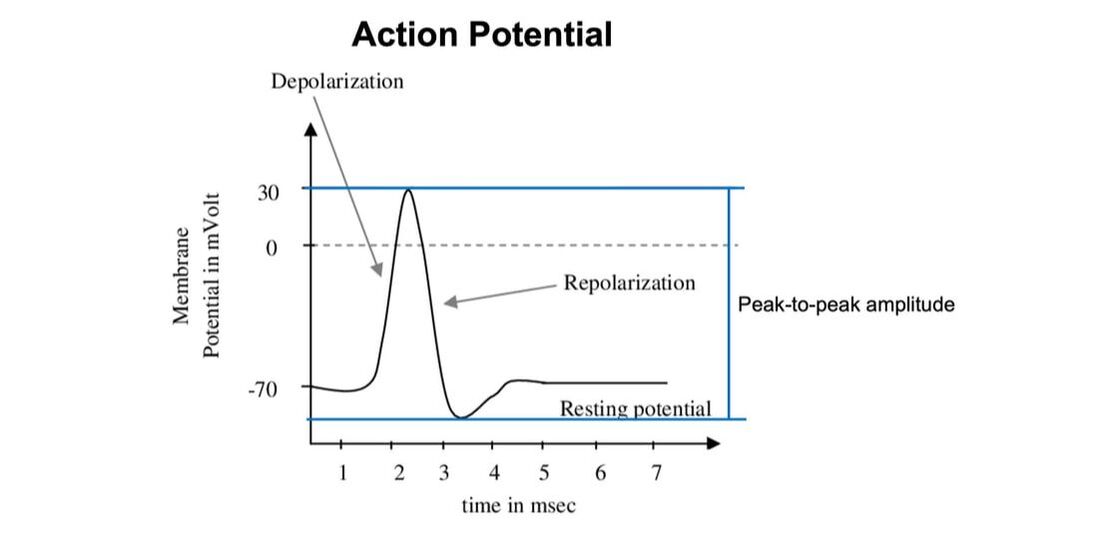 The first is obtained using needle electrodes to record from various individual motor units. The second can be recorded using surface EMG, and is obtained by maximally stimulating the muscle’s nerve to activate all motor units. Once you have both, you divide the action potential amplitude of the total motor unit pool by that of the average single motor unit—then voila, you have an estimated motor unit number! 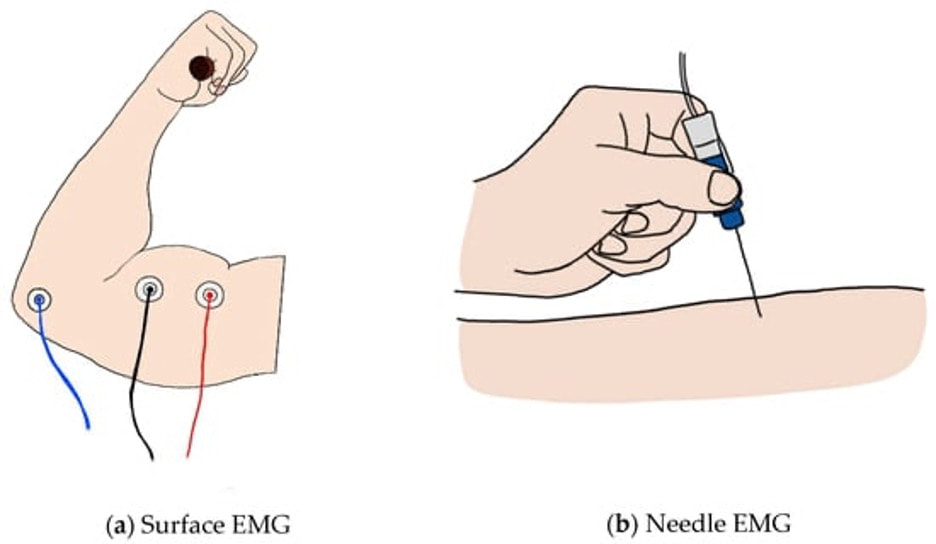 From: https://encyclopedia.pub/entry/7298 A 2005 study by McNeil and colleagues from the University of Western Ontario compared motor unit number in the tibialis anterior muscle (the muscle along your shin, pictured below) between three age groups: 25 years, 65 years, and 80 years old. From 25 to 65 years, they detected a 40% decrease in the number of motor units. That number decreased a further 33% when going from 65 to 80 years. 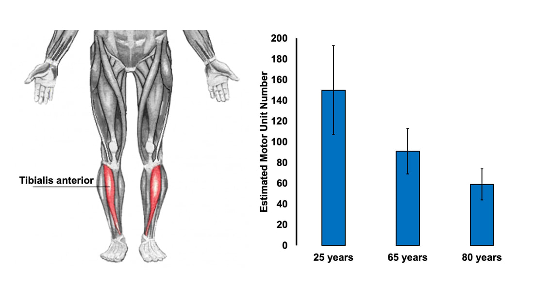 Why do we lose motor units with age and what happens with the remaining muscle fibres?Motor neurons die due to a combination of factors. Put simply, we experience damage and modifications to our DNA throughout life. Eventually, the accumulation of those DNA stresses causes cells to die, including motor neurons. This isn’t all doom and gloom, though—researchers have noted impressive ways in which the body compensates for motor unit loss. Despite observing a 40% reduction in motor unit number from ~25 to 65 years of age, the study above did not observe any differences in maximum muscle strength between those two age groups. How can this be? The easiest answer may be that the remaining motor neurons are sufficiently reinnervating the muscle fibres that get left behind. In that case, we could get a decrease in the number of motor units without a loss in the number of muscle fibres. In other words, the number of muscle fibres within a motor unit increases while the total number of motor units declines. 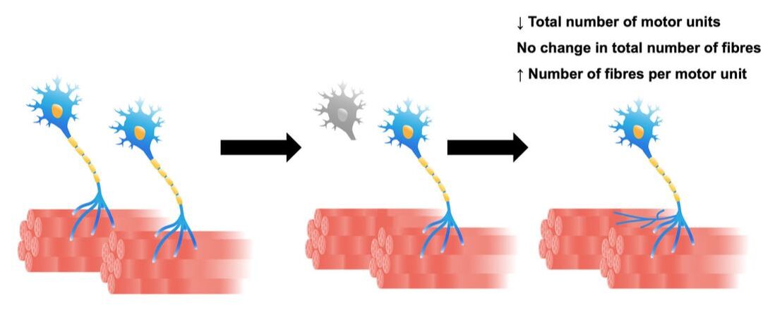 This phenomenon would preserve strength, but could still have disadvantages. With a smaller number of motor units, there would be a less gradual recruitment of muscle fibres. Instead of steady recruitment that allows for fine-tuned, smooth movements, the recruitment becomes more like a staircase, producing jerkier movements. This staircase-shaped muscle fibre recruitment is one reason you might find your movements become less stable as you age. 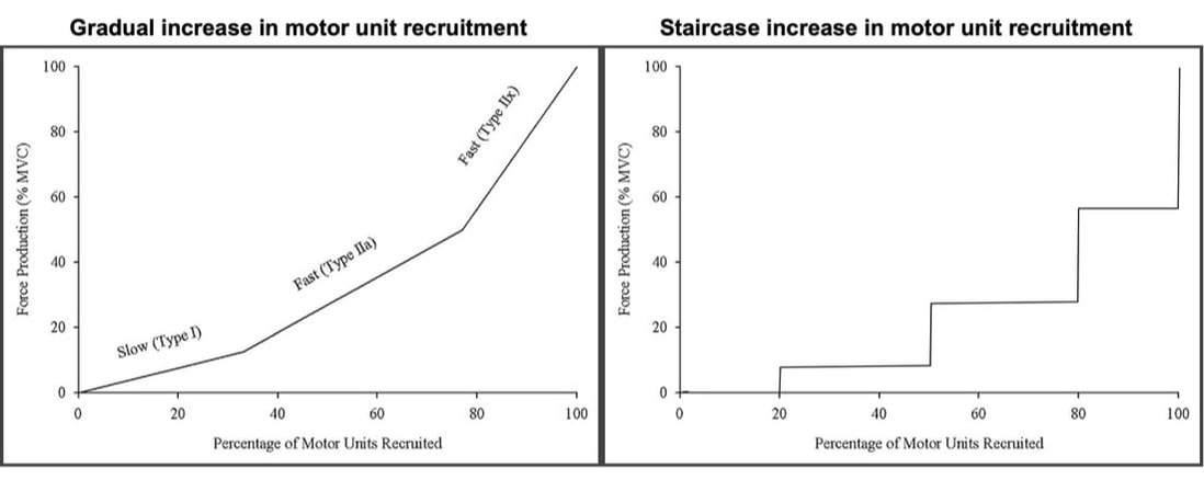 Figure on the left is from: https://pubmed.ncbi.nlm.nih.gov/15794706/. The figure on the right was not created from any data and is purely for visual comparison. But I also mentioned earlier that not all fibres are reinnervated after their motor neuron dies. Can the body compensate for that? It turns out, yes! The fibres that aren’t lost with alpha motor neuron death retain a remarkable ability to compensate for those that are lost. A study in 2008 by Frontera and colleagues tracked a group of older men for twelve years—from about age 70 to 80. They obtained measurements of strength and size in the quadricep muscles of these men, and (via muscle biopsies) in the muscle fibres that comprise those muscles. As expected, these men experienced a 6% decrease in quadricep muscle size, and 22-37% decreases in quadricep muscle strength after 12 years. 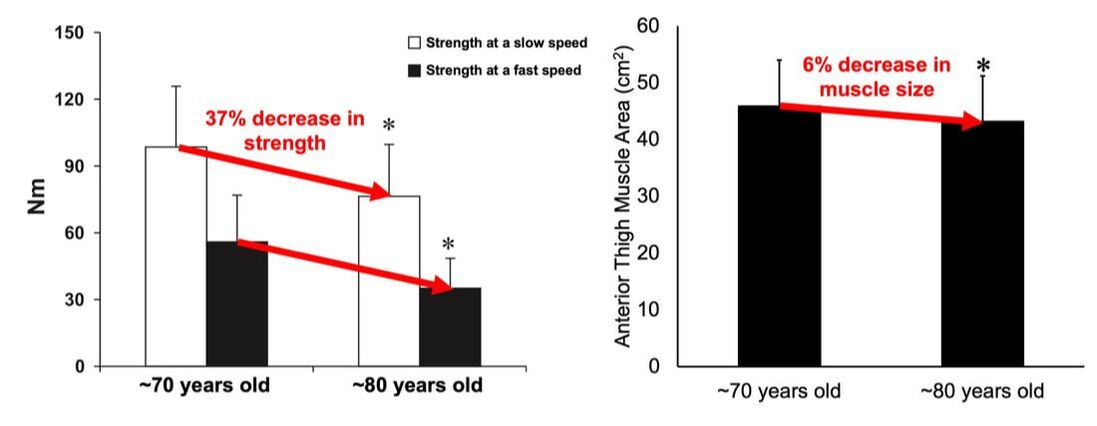 The single muscle fibres, remarkably, did not show the same trend. They saw no significant differences in fibre size or force across the 12 years. Some fibres on average even trended toward being larger and stronger. In other words, the remaining muscle fibres really try to pick up the slack of those that are lost with age. 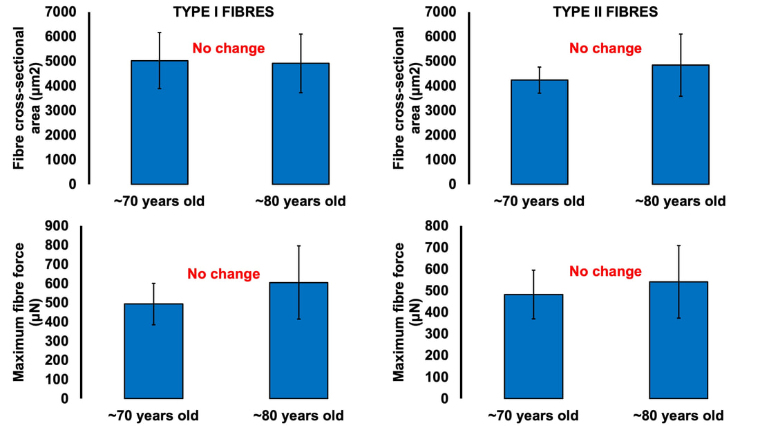 ConclusionOverall, picture motor unit loss with age using the following (a bit weird, but bear with me) analogy: You and your friend are each trying to carry an armload of rocks from Point A to Point B. Halfway there, your friend trips and drops all their rocks, and they can’t get back up. You try to compensate for this by picking up all your friend’s rocks, too. You’re able to fit most of their rocks in your arms along with your own, but don’t have quite enough space and leave some behind. You and your friend are motor neurons, the rocks are muscle fibres, and going from Point A to Point B is aging. While you may retain about the same number of muscle fibres as you age, you have fewer motor neurons working to carry them.  AUTHOR’S NOTE: I’m currently studying for my PhD candidacy exam. That leaves me with less writing time, so of course I had to turn my studying into an outlet to still write these knowledge translation articles. I figure they can help both myself and you (the reader) learn from the topics I’m studying. With that, this is the first of four knowledge translations (one for each section of my exam) that I’ll be writing over the next few months. Hopefully they’re interesting!
References McNeil CJ, Doherty TJ, Stashuk DW, Rice CL. Motor unit number estimates in the tibialis anterior muscle of young, old, and very old men. Muscle Nerve. 2005 Apr;31(4):461-7. doi: 10.1002/mus.20276. PMID: 15685623. Frontera WR, Reid KF, Phillips EM, Krivickas LS, Hughes VA, Roubenoff R, Fielding RA. Muscle fiber size and function in elderly humans: a longitudinal study. J Appl Physiol (1985). 2008 Aug;105(2):637-42. doi: 10.1152/japplphysiol.90332.2008. Epub 2008 Jun 12. PMID: 18556434; PMCID: PMC2519941.
0 Comments
Your comment will be posted after it is approved.
Leave a Reply. |
AuthorAvery Hinks Archives
September 2023
Categories |
 RSS Feed
